Hello! Welcome back!
This week, I have been repeating experiments I have previously done several times in order to either eliminate or account for an anomaly in my last round of gram-negative PCR amplifications. In initial testing, the primers I designed had amplified all 15 of the species chosen for my study. Multiple rounds of testing had positive post-PCR presence of DNA in my gels, but in a later phase of testing two of the gram-negatives did not amplify during PCR.
I am not certain at all why this occurred; there are a myriad of reasons which could account for the anomaly, including human error. So I did a new round of DNA extractions this week, ran some gels, and verified if DNA was present. All of the tested species were positive for DNA.
I subsequently banked the samples until next week, when I will be able to get some time on the thermocycler. Results of the PCR to follow on next week's blog.
Thanks for checking in with me.
Photo: Gel samples of 9 gram negative bacterial species showing DNA presence.
Translate
Thursday, October 22, 2015
Wednesday, October 14, 2015
Semester 4-Week 6-This Semester's Research Proposal
Hello! Welcome back. Since we have research proposals due this week, I am including mine here as my blog post. Please note that as my project is wrapping up, this submission does not include the normal hypothesis/projected results format that a proposal normally would. Instead, this is a preliminary version of my project abstract. Thanks for checking in this week-I appreciate your patronage!
Abstract
Abstract
Since the complete genetic
sequencing of the Escherichia coli genome
in 1997 (Blattner, et.al), variations of the 16s ribosomal gene have been found
conserved among different bacterial species. As a result, 16s ribosomal DNA
sequencing has become an integral part of bacterial identification in the
modern laboratory (Janda & Abbot, 2007). The initial step of the
identification process requires extraction of DNA, which must then be amplified
using a polymerase chain reaction (PCR) so it can be sequenced. Primers
specific to certain regions of the 16s gene are utilized to replicate the
bacterial DNA during the PCR process so that the resultant DNA can be sequenced
and correctly identified (Mao et. al 2012). Because PCR amplification is in widespread
laboratory use, knowledge of DNA extraction and PCR techniques is an
essential component of the student learner’s skill set. This study identified and
designed simple laboratory protocols for use in introducing the Phoenix College
student learner to elementary DNA identification technology. The protocols are
intended to accompany the standard clinical format of culturing both known and
unknown bacteria. The study identified electrophoresis gel techniques,
extraction protocols for gram-negative and gram positive bacterial species, a
set of universal 16s ribosomal primers that can be used to identify fifteen
gram-negative and gram-positive bacterial species available in the Phoenix
College Biosciences laboratory, and a congruent PCR amplification protocol that
can be adapted and incorporated into existing instructor curricula.
References:
References: Blattner, R.,
Plunkett III, G., Bloch, C., Perna, N., Burland, V., Riley, M., Collado-Vides,
J., & Glasner, J. (1997). The complete genome sequence of escherichia coli
k-12. Science, 277, 1453-1462. doi:
10.1126/science.277.5331.1453.
Cattelino, P. (2014)
Identification and application of universal 16s rRNA ribosomal primers. Phoenix College Student Paper. Pps.
1-16.
Frank, J., Reich, C.,
Sharma, S., Weisbaum, J., Wilson, B., & Olsen, G. (2008). Critical
evaluation of two primers commonly used for amplification of bacterial 16s rRNA
genes. 74(8), 2461-2470.
Janda, J., & Abbot,
S. (2007). 16s rRNA gene sequencing for bacterial identification in the
diagnostic laboratory: Pluses, perils, and pitfalls. Journal of Clinical
Microbiology, 45(9), 2761-2764.
Mao, D., Zhou, Q., Chen,
C., & Quan, Z. (2012). Coverage evaluation of universal bacterial primers
using the metagenomic datasets. BMC Microbiology, 12(66),
Retrieved from www.biomedcentral.com/1471-2180/12/66.
Marchesi, J., Soto, T., Weightman, A., Martin, T., Fry, J.,
Hiom, S., & Wade, W. (1997). Design and evaluation of useful bacterium-specific
PCR primers that amplify genes coding for bacterial 16s rRNA. Applied and
Environmental Microbiology, 64(2), 795-799.
PS: Just because I LOVE space exploration and NASA, here is a link to NASA's news page and an image of one of Jupiter's moons, Io, for your enjoyment.
This global view of Jupiter’s moon, Io, was obtained during the tenth orbit of Jupiter by NASA’s Galileo spacecraft. Credit: NASA
Friday, October 9, 2015
Semester 4-Week 5: Extraction Protocols
Hello! Welcome back.
I have had a couple of people ask me for bacterial DNA extraction protocols this semester, so I am going to publish them on this blog this week. One is an SDS thermal extraction for gram-negatives, and the second is a quick boil method for gram-positives. Feel free to copy and paste and make them your own!
Protocol#1
Spin down 500 µl of culture in a 1.5 mL tube at 12,000 rpm for 3 minutes. Repeat to increase size of base sample; total volume of sample spun down not to exceed 4.5 mL. Re-suspend cells in 0.3 mL TBS and 15 µL of 10% SDS (0.5% SDS final concentration). Incubate the solution in 55°C heat block for 10 minutes. Cool down to room temperature. Add 5 µl proteinase K solution @ dilution ratio of 5 mg powdered proteinase K per 250 µl of sterile molecular-biology grade water. Vortex ~20-30 seconds. Incubate the sample on ice for 5 minutes. Spin the sample at 12,000 rpm for 3 minutes. Verify the pellet is not loose. Transfer supernatant to a clean 1.5 mL tube. Add 300 µL of room temperature isopropanol. Gently mix by inversion until the chromosomal DNA threads to form a visible mass. Centrifuge the DNA at 12,000 rpm for 2 minutes. Carefully pour off the supernatant, use a clean pipette to remove any remaining supernatant, and add 300 µL room temperature 70% ethanol. Invert several times to wash the DNA pellet. Centrifuge at 12,000 rpm for 2 minutes. Drain off the supernatant, use a clean pipette to remove any remaining supernatant, and allow the DNA pellet to air dry under hood for 15-20 minutes. Re-suspend the DNA pellet in 50 µL sterile molecular-biology grade water.
Protocol #2:
Incubate culture for 24 hours @ 37°C. Pellet 500 µl of culture at 12,000 x g for 3 minutes. Re -suspend pellet in 500 µl sterile water using a 30 second vortex. Incubate sample for 10 minutes @ 100°C. Pellet @ 12,000 g for 3 minutes. Pour off supernatant into 1.5 ml Eppe tube and discard pellet.
Until next week, please enjoy this National Geographic article about Homo naledi, a human ancestor recently discovered in a South African cave (artist rendition below):
Photo credit: National Geographic Magazine, September 10, 2015
I have had a couple of people ask me for bacterial DNA extraction protocols this semester, so I am going to publish them on this blog this week. One is an SDS thermal extraction for gram-negatives, and the second is a quick boil method for gram-positives. Feel free to copy and paste and make them your own!
Protocol#1
Spin down 500 µl of culture in a 1.5 mL tube at 12,000 rpm for 3 minutes. Repeat to increase size of base sample; total volume of sample spun down not to exceed 4.5 mL. Re-suspend cells in 0.3 mL TBS and 15 µL of 10% SDS (0.5% SDS final concentration). Incubate the solution in 55°C heat block for 10 minutes. Cool down to room temperature. Add 5 µl proteinase K solution @ dilution ratio of 5 mg powdered proteinase K per 250 µl of sterile molecular-biology grade water. Vortex ~20-30 seconds. Incubate the sample on ice for 5 minutes. Spin the sample at 12,000 rpm for 3 minutes. Verify the pellet is not loose. Transfer supernatant to a clean 1.5 mL tube. Add 300 µL of room temperature isopropanol. Gently mix by inversion until the chromosomal DNA threads to form a visible mass. Centrifuge the DNA at 12,000 rpm for 2 minutes. Carefully pour off the supernatant, use a clean pipette to remove any remaining supernatant, and add 300 µL room temperature 70% ethanol. Invert several times to wash the DNA pellet. Centrifuge at 12,000 rpm for 2 minutes. Drain off the supernatant, use a clean pipette to remove any remaining supernatant, and allow the DNA pellet to air dry under hood for 15-20 minutes. Re-suspend the DNA pellet in 50 µL sterile molecular-biology grade water.
Protocol #2:
Incubate culture for 24 hours @ 37°C. Pellet 500 µl of culture at 12,000 x g for 3 minutes. Re -suspend pellet in 500 µl sterile water using a 30 second vortex. Incubate sample for 10 minutes @ 100°C. Pellet @ 12,000 g for 3 minutes. Pour off supernatant into 1.5 ml Eppe tube and discard pellet.
Until next week, please enjoy this National Geographic article about Homo naledi, a human ancestor recently discovered in a South African cave (artist rendition below):
Friday, October 2, 2015
Semester 4-Week 4
Hello! Welcome back.
I am still compiling my data into a comprehensible paper, so there are no new experiments yet to report. I will only be doing lab work this semester if I run into incomplete notes on data in my lab book. If that happens, then I will need to re-run that particular experiment to complete my data set.
I was going to explain primer design on this blog, but instead I found an online resource that explains it more thoroughly than I ever could, so I have included a link to it here: http://www.cybertory.org/exercises/primerDesign/ . The lesson from Cybertory.org is one of the simplest and on-point explanations I have ever found and is a great introduction to how primers work.
I am also including a link here from Bioinformatics.org that will assist you in designing your own primers. Of course, if you would like to manually reverse-complement your DNA strands, go for it, but I prefer an online generator to reduce the chance of transposing a nucleotide and rendering my primer useless. With the Bioinformatics link, you just type in the gene sequence you are targeting, then click the function you need (reverse/complement, etc.), and your new primer sequence is immediately generated.
There are two other great resources for finding gene sequences and/or designing primers. One of them is the NBCI database, which contains an extensive listing of genomes. I have included a Youtube tutorial here (and below this post) which explains how to use the database. The home page pictured in the tutorial has changed since it was posted, but the process is still the same (the format is just slightly different). Once you find your strain of bacteria, you can simply click the "Get Primers" button and they will be provided for you, along with useful information such as the region of the gene being targeted. You can also select your parameters, as far as base pair size, by using the editing tool provided.
Another good resource is the Straininfo.net database located here. This database is particularly useful if you are using a patented strain of bacteria, are targeting the 16s rRNA gene, and are unsure of the original source strain. This was the case with my E. coli. Simply go to the link, then type your strain in the search box (for example, ATCC 4157). This will return a page that displays an overview of the strains available, and yours should be highlighted if it is in the database. After verifying that yours is available by finding the highlighted area, just navigate below the strain listing and click the "SeqRank" icon. On the bottom left of the page will be the recommended 16s rRNA sequence. Just copy and paste, edit the weird formatting so that you have a single continuous nucleotide sequence, and you are ready to analyze your gene for potential areas to target with primers.
One final word on primer design. There are other online tools available to help you design your own set that will allow you to load in genes and scan for similarites. In my case, however, I resorted to simply downloading all of the 16s rRNA sequences for the bacteria used in my study into a single Word document. Then, I looked for correlations between the genes by using the "Ctrl F" function. It was painstaking, but fun.
I hope this helps with your own primer design. See you next blog!
Wednesday, September 23, 2015
Semester 4-Week 3
Hello! Welcome back.
As of this blog, I am working on the initial phase of my final project paper. This phase deals with writing the "Material and Methods" section, as well as a brief summary of the outcomes of the various DNA extraction protocols I tested. I have also collected the protocols together into a series of appendices that will be included in the final result.
In between this, I have been doing an intense amount of studying. For some reason, my classes this semester require a lot of self-driven learning and repeated in-class exams. This is a bit of a change from previous semesters, so I have had to adjust my study habits accordingly.
For the science portion of this week's blog, I looked at infection rates for malaria and potential treatments. Malaria infects human erythrocytes when the organism Plasmodium falciparum is transmitted into the bloodstream via the bite of the Anopheles mosquito. While parasitic malaria infection has a wide range of symptoms, and typically presents as chills, fever, sweating, and vomiting, it is potentially fatal, particularly in those with a weakened or underdeveloped immune system. The primary method of death involves the disruption of blood flow to vital organs. According to a 2015 report by the World Health Organization, infection rates have declined by 47% globally since the year 2000, yet severe malarial cases still kill an estimated 584,000 people per year, primarily African children.
Drug-resistant strains of malaria have mutated and proven difficult to treat with conventional methods. However, according to a study by Nobel laureate Sidney Altman recently reported on in Medical News Today by Catharine Paddock, malaria cases can be treated by disrupting the gene expression of P. falciparum by RNA. Researchers used a tool called a morpholino oligomer to disrupt expression, which resulted in slower development rates of the parasite. The lower development rates delayed the onset of full-blown infection. This new technique is a promising development in the treatment of drug-resistant strains of the infection.
I am including links to a number of articles, and the abstract for the published study. Please enjoy them!
Photo credit: National Geographic. Image: The female Anopheles mosquito is the only mosquito species that carries the malaria parasite P. falciparum.
Centers for Disease Control: http://www.cdc.gov/malaria/about/disease.html
Medical News Today article: http://www.medicalnewstoday.com/articles/299207.php
World Health Organization: http://www.who.int/mediacentre/factsheets/fs094/en/
Source for the journal publication about gene therapy:
Targeting protein translation, RNA splicing, and degradation by morpholino-based conjugates in Plasmodium falciparum, Aprajita Garg et al., Proceedings of the National Academy of Sciences, doi:10.1073/pnas.1515864112, published online 8 September 2015
Link to the abstract: http://www.pnas.org/content/112/38/11935.abstract
Friday, September 18, 2015
Semester 4-Week 2
Hello! Welcome back.
This week was spent primarily focused on academics, but as of this writing I have also begun my final paper about this project. This is probably going to take some time to do, as I have to sift through all of my notes from the last three semesters. I haven't always been as organized with my lab notes as I have learned to be, so I am anticipating that some of my data may be incomplete. If so, I will have to repeat portions of my project several times in order to verify the accuracy of the data that I do have.
Live and learn. Good note-keeping=the best possible accuracy.
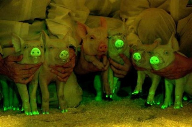
Also this week, I spent a little time enjoying the following articles about GFP, or Green Fluorescent Protein. This particular protein was discovered in a species of jellyfish, Aequorea victoria, in the late 20th century and subsequently has proven useful for tracking protein production and gene expression in a number of animal species. The protein glows green under light in the blue-ultraviolet range, and while originally sourced from the jellyfish species, this protein was cloned in 1994 and thereafter became commercially available.
Please peruse the links. Also, please take a look at the pics; the animals shown have been altered with GFP.
http://www.conncoll.edu/ccacad/zimmer/GFP-ww/GFP-1.htm
http://www.uniprot.org/uniprot/P42212
Photo credits: www.buzzfeed.com
Wednesday, September 9, 2015
Semester Four, Week 1-Welcome Back!
Hello! Welcome back to my blog!
I am so glad to be back this semester, for my final S-STEM enrollment. I have hit the home stretch of this amazing experience, and shortly after it ends I will be heading off to ASU to complete my degree.
For the rest of this semester, though, I will be focused on compiling my project data into a coherent narrative that presents the work I have done in this program. I will still be setting up experiments, where necessary, to collect additional data, but most of my time this session you will likely find me buried in a notebook or on my laptop.
Feel free, though, to stop by and say hello. And if you are new to S-STEM, don't hesitate to contact me with any questions you have about anything. I'm here to help whenever I can.
See you in the lab!
Photo credit: FLICKR/NIAID
Here, a fractured Vero cell provides a view into a vacuole, which houses a growing cluster of Coxiella burnetii. This form of bacteria can cause Q fever, a bacterial infection that affects the heart, liver, lungs and other organs.
Subscribe to:
Posts (Atom)